Shave removal
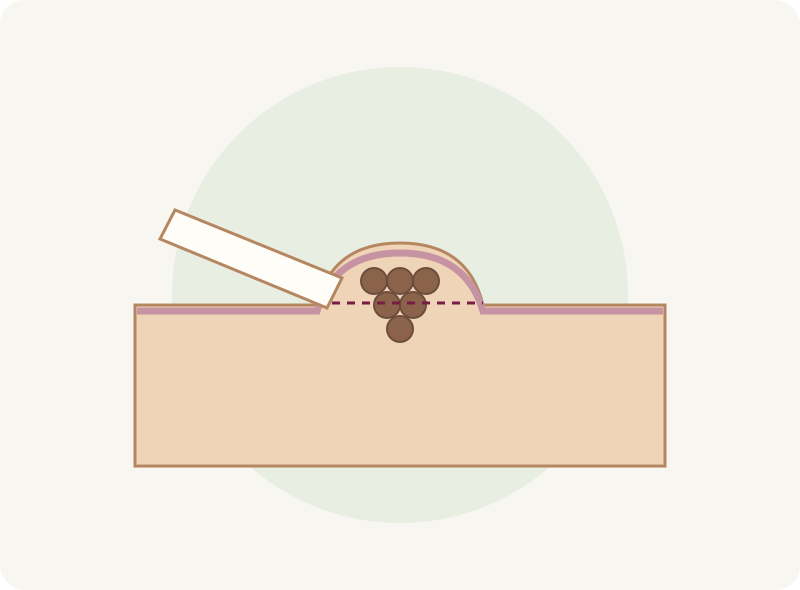
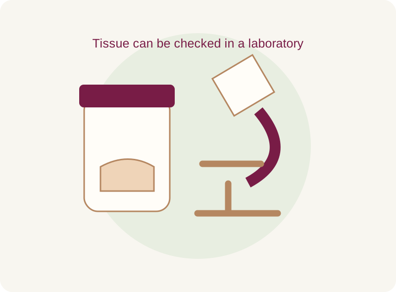
Selected raised, benign moles may be shaved under local anaesthetic. Tissue can be sent for examination. Some cells may remain, so regrowth is possible.
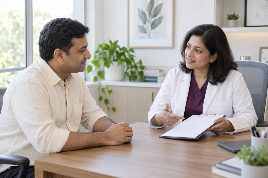
Whether a mole catches on clothing or has changed, an examination comes first. Understand what it is, whether removal is needed and how the area may heal.
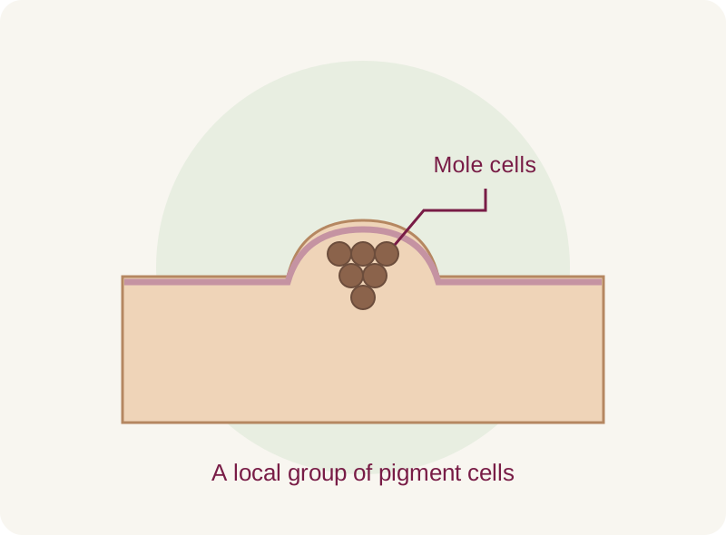
Moles form from groups of pigment-producing cells. They may be flat or raised and range from skin-coloured to dark brown. Most are harmless and can be left alone.
Removal may be considered for repeated irritation, a cosmetic preference or a concern that needs tissue testing. A dermatologist checks the lesion before choosing a method.
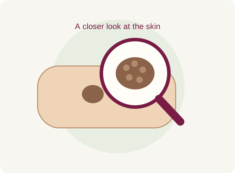
Book an assessment for changes in shape, colour or size, or a spot that looks different from your other moles. Small spots can also need checking.
Mention these symptoms, even if you think clothing or shaving caused them. Do not cover up a persistent change with a cosmetic treatment.
Tell the dermatologist about previous skin cancers, family history and when the mark appeared. Older photographs can help explain changes.
Selected raised, benign moles may be shaved under local anaesthetic. Tissue can be sent for examination. Some cells may remain, so regrowth is possible.
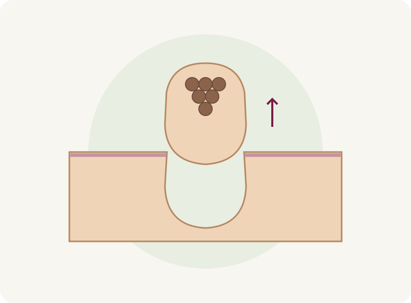
The lesion is cut out to the required depth, often with stitches to close the skin. This can provide a fuller tissue sample when diagnosis requires it.
Observation is also an option. A suspicious lesion needs the right biopsy or referral; laser, freezing or cautery should not replace a needed tissue sample.
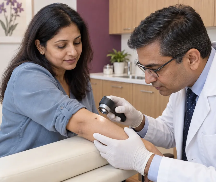
The clinician may use a dermatoscope and discuss observation, removal or further investigation.
Discuss the likely scar, tissue testing, medicines, allergies and healing history. Do not stop prescribed medicines without advice.
Local anaesthetic numbs the area; the injection may sting. The clinician uses the agreed method and controls any bleeding.
Stitches or a dressing may be needed. You receive a wound-care plan and any follow-up arrangements.
If tissue was tested, ask when and how you will receive the report. Further care depends on the findings.
Keep any wound-review or stitch-removal appointment. A mole that returns should be examined again.
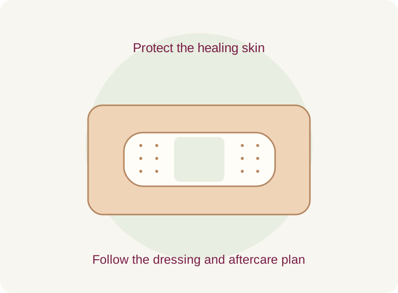
Clean gently and use the advised ointment and dressing. Avoid picking or soaking the wound while it heals; ask when to resume sport and swimming.
A scar, temporary colour change, soreness or bleeding can occur. Healing varies with the site and method. Once healed, protect exposed skin from the sun.
Increasing pain, warmth, swelling, discharge or persistent bleeding needs medical advice. Report a new growth or pigment appearing in the treated area.
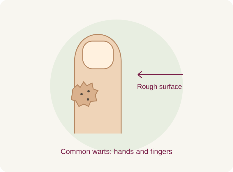
Viral warts are different from pigment-cell moles. Assessment helps choose the right approach.
Explore wart care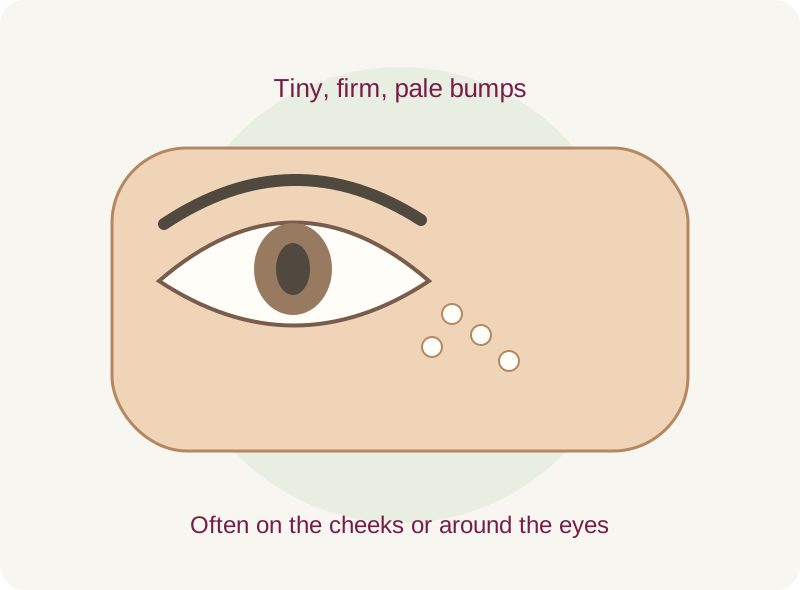
Tiny white keratin cysts are another kind of skin bump, often managed with simple care.
Explore miliaVerified Google review excerpts about consultations at Layers. The condition is not specified; these are not mole-removal results.
“The doctors listened carefully to my concerns and explained the treatment in a clear and professional manner.”
Consultation experience; condition not specified.
“She explained the treatment clearly and made me feel comfortable throughout the consultation.”
Consultation experience; condition not specified.
“The doctor took the time to understand my skin concerns and explained the treatment plan clearly, without rushing.”
Consultation experience; condition not specified.
“Very patient ,humble and polite to answer every single query and build confidence.”
Consultation experience; condition not specified.
“The doctors explained everything clearly and made me feel comfortable and confident.”
Consultation experience; condition not specified.
“The doctor took the time to listen to my concerns patiently and explained the treatment options clearly.”
Consultation experience; condition not specified.
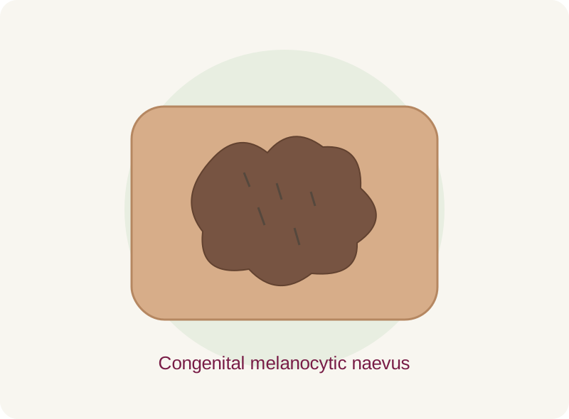
A mole present from birth may need a monitoring or specialist plan based on its size, location and changes. Removing its colour is not the same as assessing the whole lesion.
Understand birthmarks



No. A benign mole that is not troublesome may be left alone. Your preference, symptoms and the examination guide the decision.
Diagnosis comes before the device. Destructive methods can leave no sample for testing and may allow regrowth. Ask why the proposed method is appropriate; a suspicious lesion needs diagnostic care.
Any removal can leave a scar or colour change. The size, location, method and your healing history affect the result. Scar-free healing cannot be guaranteed.
Shave removal often heals without stitches. Excision commonly needs closure with stitches. The clinician will explain what applies to your lesion.
Local anaesthetic is commonly used. The injection can sting and the area may feel sore afterwards. Tell the clinician if you feel pain during removal.
Tissue examination is important when the diagnosis needs confirmation. Before removal, ask whether a sample will be sent and how the findings will be shared.
Yes, especially after partial removal. Do not assume a recurring mark is harmless; arrange another examination.
Do not cut, burn or apply acids to a mole at home. This can cause a wound or scar and delay diagnosis of a concerning lesion.
Get advice on examination, removal and follow-up for the mark that concerns you.
Prefer a call? +91 9000 572727