
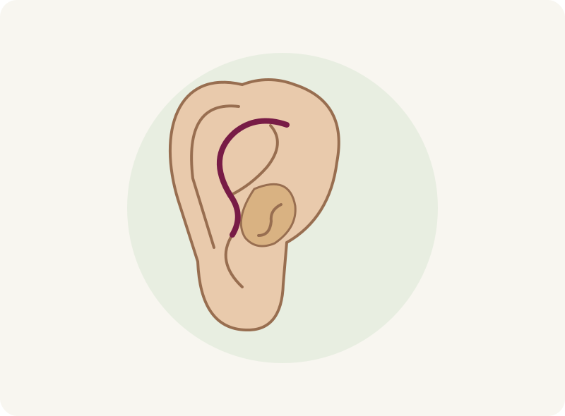
The inner fold
A less-defined cartilage fold can contribute to a prominent ear. Reshaping or supporting the fold may be part of the plan.
Otoplasty changes the shape or position of the outer ear. Learn what can be adjusted, how surgery is planned and how to protect your ears while they heal.
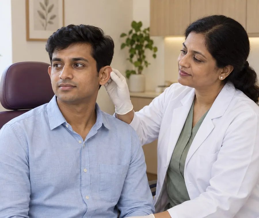
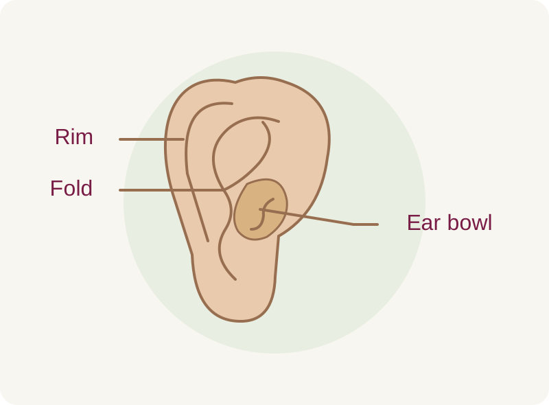
Otoplasty reshapes the outer ear, often to reduce how far it projects from the head. The surgeon may adjust cartilage folds, the ear bowl or their relationship to the head. The plan can involve one or both ears.
The aim is a shape that fits your features. This operation does not improve hearing, and perfect left-to-right symmetry is not a realistic promise. Hearing symptoms need their own assessment.
A less-defined cartilage fold can contribute to a prominent ear. Reshaping or supporting the fold may be part of the plan.
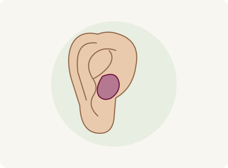
The bowl’s depth and shape also affect projection. The surgeon assesses it separately rather than applying one technique to every ear.
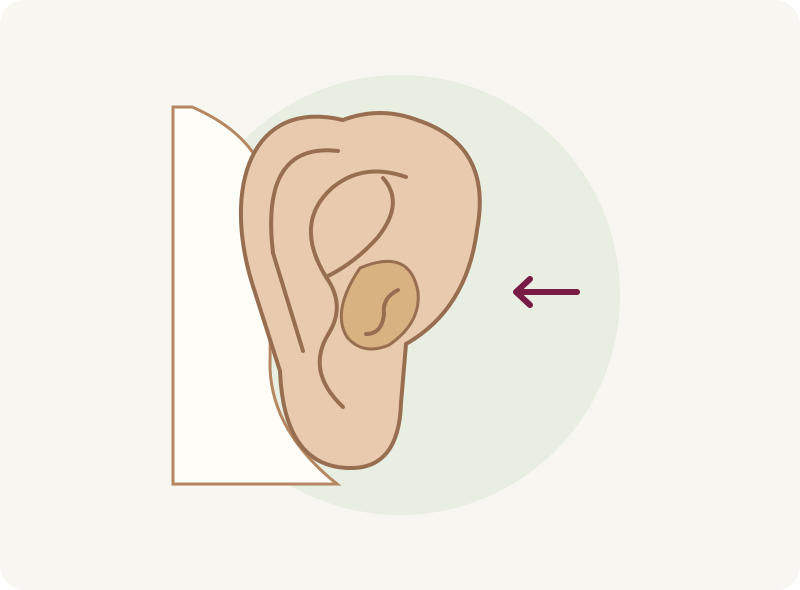
Ear-to-head position and the balance between the two sides matter. The aim is a natural contour rather than an ear pulled excessively flat.
Explain which aspect of your ears bothers you and how long you have considered surgery. The choice should be yours, with realistic expectations and time to decide.
The surgeon examines size, shape, symmetry and previous scars. For a child, ear development, readiness and the child’s own wishes are part of the decision.
Discuss ear infections, medicines, nicotine use, past surgery and unusual scarring. You need to be able to protect the ears and follow the dressing and review plan.
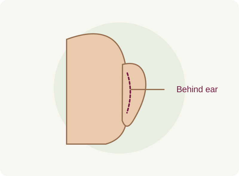
Incisions are commonly placed behind the ear. Some plans use access within a front fold. The location depends on which cartilage or tissue needs adjustment.
Internal stitches can support the reshaped cartilage. Skin incisions are closed separately. Scars remain, even when placed in a less visible crease, and the surgeon should explain any risk of noticeable or raised scarring.
The surgeon examines your ears and may take photographs. Discuss which structures would be adjusted, likely scars and the limits of symmetry.
Confirm anaesthesia, the surgical setting, transport and follow-up. Follow your preparation plan; any change to prescribed medicines needs the prescribing clinician’s agreement.
Local anaesthesia with sedation or general anaesthesia may be used. The surgeon makes the agreed incision and adjusts the selected cartilage or soft tissue.
Stitches secure the planned contour and close the wound. Dressings support the ears during early healing, and the team checks your recovery before discharge.
Keep dressings in place as directed. Contact the team if they slip, become wet or cause increasing pain; avoid removing or tightening them yourself.
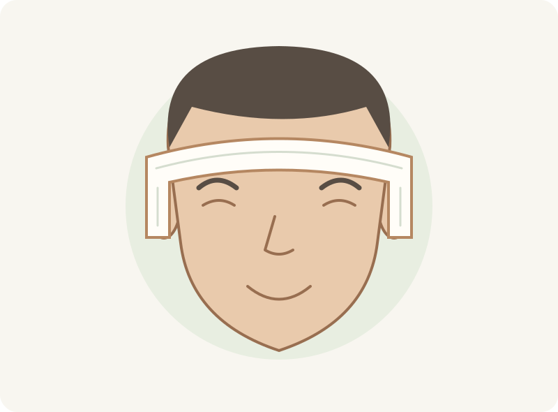
Follow instructions about sleeping, hair washing, glasses, headphones and headbands. Return to exercise and contact sports only when cleared, and attend scheduled reviews.
The ears may feel sore, itchy, swollen or bruised at first. Sensation can change temporarily. Ask the team how to manage discomfort and when dressings or external stitches will be checked.
Work, school and sports have different demands on healing ears. Plan around the advice for your operation, including protection during sleep and activities. Early appearance can change as swelling settles.
Bleeding under the skin, infection, poor wound healing, persistent pain or altered sensation can occur. Anaesthetic risks and reactions to dressings or stitches also need discussion.
Scars may remain noticeable or become raised. Internal stitches can sometimes irritate the skin or need further attention.
Uneven ears, an overly flat appearance, contour irregularities or some return of prominence are possible. Revision may be needed and has its own limits.
These Google reviews describe general consultations and care at Layers. They are not otoplasty outcome reports.
“The doctors listened carefully to my concerns and explained the treatment in a clear and professional manner.”
Consultation experience; condition not specified.
“She explained the treatment clearly and made me feel comfortable throughout the consultation.”
Consultation experience; condition not specified.
“The doctor took the time to understand my skin concerns and explained the treatment plan clearly, without rushing.”
Consultation experience; condition not specified.
“Very patient ,humble and polite to answer every single query and build confidence.”
Consultation experience; condition not specified.
“The doctors explained everything clearly and made me feel comfortable and confident.”
Consultation experience; condition not specified.
“The doctor took the time to listen to my concerns patiently and explained the treatment options clearly.”
Consultation experience; condition not specified.














No. It changes the outer ear’s appearance. Ear pain, hearing loss or recurrent infections should be assessed separately.
Sometimes. Both ears are assessed because their relationship affects the plan. The surgeon may recommend one-sided or two-sided surgery.
Reducing projection and reducing ear size are different changes. Explain your concern so the surgeon can discuss the appropriate operation.
It may be considered once the ears are sufficiently developed and the child is ready for surgery and aftercare. A specialist should assess the child’s needs and wishes.
Ear moulding may help some newborn ear-shape concerns when assessed very early. It is not a substitute for adult cartilage surgery. Ask a paediatric ear specialist promptly if this concerns a baby.
A protective headband may be part of the aftercare plan after the initial dressing. The type, fit and duration should come from your surgical team.
Some loss of correction or change over time is possible. Protecting healing ears and attending review matter, but no procedure guarantees a fixed shape for life.
Ask about the surgeon’s relevant experience, incision location, anaesthesia, aftercare support and how concerns or revision would be managed.
Discuss shape, recovery and the change that matters to you before deciding on surgery.
Prefer a call? +91 9000 572727